Tools of the trade, explained
Zebrafish
Zebrafish (Danio rerio) are tropical fish. Their eggs are small, optically transparent, and easily accessible. This enables us to monitor biological events in real time at high resolution, in a manner not easily achieved in other systems. Zebrafish develop rapidly, so they are perfectly suited to embryological manipulations and genome-editing. Since many genes are conserved among vertebrates, insights gained from functional studies in zebrafish are relevant to human biology and disease. Their genetic similarity to humans allows us to carry out large scale genetic and drug discovery studies.

Myelin
Myelin is the lipid-rich membrane of specialised glial cells, which wraps around axons — the cable-like projections that neurons extend to communicate with other cells — to insulate them, like electrical tape around a wire. In the brain and spinal cord, myelin is made by oligodendrocytes.
Myelinated axons constitute about half the volume of our brain and spinal cord, and are the information superhighways of our nervous system. Myelination starts around birth and continues well into adult life. It is dynamically regulated, including by our brain activity, and this represents a way in which the function of our neural circuits can be modified.
Disruption to the formation, function or healthy maintenance of myelinated axons is associated with many human diseases throughout the life-course, from neurodevelopmental conditions through to neurodegenerative disorders, including the demyelinating disease multiple sclerosis (MS). We aim to understand myelinated axon biology to gain insight into brain formation and function and to inform strategies to treat disease.

Genetic Screens
Zebrafish can easily be used to carry out forward genetic screens — also known as gene discovery screens. In a forward genetic screen, mutations are randomly induced in the DNA of adult fish. We then screen through their offspring, looking for abnormal myelin phenotypes. Once we find such a phenotype, we can sequence the DNA of the fish to find which gene was affected by the mutation, and thus discover new genes which affect the function of myelin. Our lab has discovered several myelin-regulating genes in this way (see Kegel et al., 2020, and Klingseisen et al., 2019).
In recent years, we have incorporated new technologies — principally those based on the CRISPR-Cas system — that allow rapid and routine generation of mutations in genes of interest to interrogate gene function in a targeted manner in vivo.

From Kegel et al. (2020)
Chemical Screens
The small size and rapid development of zebrafish means that they can easily be used to carry out chemical biology screens, simply by adding the compound to be tested to the embryonic medium in which the zebrafish swim freely. We can use such screens to identify compounds that affect various aspects of myelinated axon formation (see Early et al., 2018).
We have established a platform to carry out fully automated drug discovery screens in zebrafish at high resolution in our UK zebrafish screening facility.

Imaging Techniques
We have taken significant time and effort to generate a suite of transgenic zebrafish lines and reagents that allow us to visualise many aspects of myelinated axon formation and function over time in the living animal, using our various microscopes.
Confocal imaging
A Zeiss LSM880 with Airyscan is the workhorse for our structural and function imaging. The Airyscan unit allows for super-resolution imaging, down to 140nm in x-y and 400nm in z. Our LSM880 has a piezo z-drive and the Airyscan FAST module, allowing for rapid volumetric imaging, ideal for structural time-lapse or functional imaging experiments.
VAST
Our Vertebrate Automated Screening Technology (VAST) zebrafish screening robot, developed by the Yanik Lab and commercialised by Union Biometrica, automatically loads larvae from 96-well plates and orients them ready for imaging. We’ve combined VAST with a high-speed spinning-disk confocal microscope for rapid imaging of hundreds of zebrafish larvae per day at sub-cellular resolution (see Early et al., 2018). Automation of image processing allows us to rapidly identify changes to myelination.
Two photon
We are in the process of purchasing a new imaging platform, including a 2-photon point scanning confocal microscope, a spinning disk confocal microscope, as well as 2-photon holographic light patterning technology. This new system will allow for advanced functional imaging and optogenetic manipulation, in combination with high resolution structural imaging.
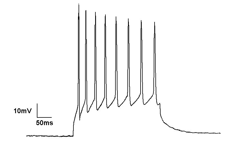
Electro-physiology
Neurons, principal cells of the nervous system, use electrical signals to communicate to each other and to their environment. Electrophysiology measures these electrical signals in different types of neurons, in health and disease. Using this technique we can detect either action potentials (electrical impulses, measured in volts) or electrical currents (measured in amperes). These values can inform us about cellular maturity level, their health status as well as molecular composition.
In our lab, we use electrophysiology to measure the speeds at which electrical impulses travel along the cell. When the protective myelin sheaths are disrupted, for example in multiple sclerosis, these electrical impulses slow down significantly. We want to know how alterations to myelin lead to abnormal neuronal signalling, and whether altering this signalling (for example, using light stimulation in genetically modified fish) would have an impact on the myelin itself.